Clinical reasoning · Emergency medicine · OSCE guide
DKA vs HHS in OSCEs: Diagnosis, Blood Gas Interpretation and Emergency Management
This is your ultimate DKA vs HHS OSCE guide: learn to distinguish diabetic ketoacidosis from hyperosmolar hyperglycaemic state, interpret the critical blood results, and explain the safest first steps in an emergency station.
The challenge? These two diabetic emergencies can look similar—but the management priorities are not.
Three OSCE traps this guide will help you avoid
- Ruling out DKA because the glucose looks almost normal.
- Starting insulin without checking potassium.
- Correcting HHS too rapidly instead of monitoring osmolality.
The OSCE.ai approach to a standout OSCE answer: Recognise the danger → Interpret the numbers → Choose the safe action → Explain your reasoning. That practical sequence—not memorising thresholds alone—is the secret to showing an examiner your best clinical thinking.
What you'll master in this guide
- Diagnosis: current DKA, HHS and mixed-crisis criteria, plus severity grading.
- Interpretation: a worked blood gas, anion gap and osmolality calculation.
- Safe management: ABCDE, fluids, potassium, insulin timing and reassessment.
- Exam practice: four examiner-style cases, model presentations and a 20-point checklist.
This guide focuses on adult patients and primarily uses the 2026 ADA Standards of Care and 2024 international hyperglycaemic crises consensus. UK JBDS differences are identified where clinically relevant. Paediatric and pregnancy-related emergencies require adapted specialist protocols.
How to read the clinical highlights
Each color is a study cue—not a measure of disease severity.
- Essential knowledgeDiagnostic criteria, biochemical thresholds, and key concepts.
- Patient safetyUrgent risks, treatment precautions, and mistakes to avoid.
- Examiner insightClinical reasoning and actions that earn OSCE marks.
1. DKA vs HHS: The 60-second OSCE comparison
Both DKA and HHS are potentially life-threatening diabetic emergencies involving insulin deficiency and metabolic disturbance.
However, their underlying physiology, clinical presentation and treatment priorities differ.
| Feature | Diabetic ketoacidosis (DKA) | Hyperosmolar hyperglycaemic state (HHS) |
|---|---|---|
| Typical onset | Hours to days | Days to a week or longer |
| Common association | Type 1 diabetes; also type 2 | More often type 2 diabetes |
| Main disturbance | Ketonaemia and metabolic acidosis | Hyperosmolality and major dehydration |
| Blood glucose | Elevated or near-normal | Usually markedly elevated |
| Ketones | Significantly elevated | Absent or mildly elevated |
| Acidosis | Present | Significant ketoacidosis absent |
| Common symptoms | Vomiting, abdominal pain, deep breathing | Extreme thirst, weakness, confusion |
| Main treatment emphasis | Fluids, potassium safety and insulin to clear ketones | Controlled rehydration, osmolality monitoring and carefully timed insulin |
High-yield OSCE distinction: DKA is primarily a problem of ketone accumulation and metabolic acidosis, whereas HHS is dominated by severe dehydration and hyperosmolality.
Neither condition should be diagnosed or treated based on glucose alone.
2. Why do DKA and HHS develop?
DKA: Insulin deficiency causes ketone production
In DKA, insufficient effective insulin and increased counter-regulatory hormones promote the breakdown of fat into free fatty acids.
The liver converts these fatty acids into ketone bodies, particularly beta-hydroxybutyrate and acetoacetate.
As ketoacids accumulate, they cause metabolic acidosis.
At the same time, increased hepatic glucose production and reduced glucose utilisation contribute to hyperglycaemia. Glucose-related osmotic diuresis causes water and electrolyte loss.
This explains the typical findings:
- Polyuria and polydipsia from osmotic diuresis.
- Vomiting and abdominal pain associated with ketoacidosis.
- Deep Kussmaul breathing as respiratory compensation for metabolic acidosis.
- Tachycardia, dehydration and potentially circulatory shock.
HHS: Residual insulin limits ketosis but not hyperglycaemia
In HHS, residual insulin is often sufficient to suppress major ketone production but insufficient to prevent severe hyperglycaemia.
Prolonged osmotic diuresis causes substantial fluid loss. As dehydration progresses, renal glucose clearance decreases, worsening hyperglycaemia and plasma hyperosmolality.
Neurological impairment may develop as the hyperosmolar state becomes more severe.
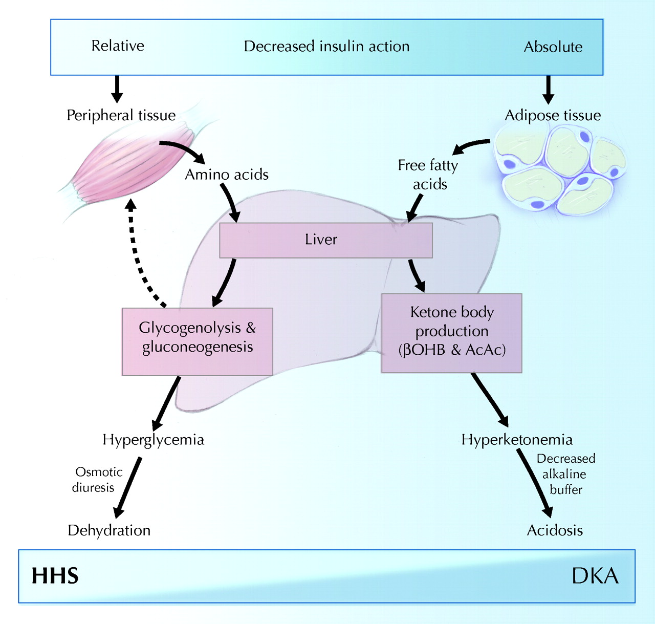
Examiner pearl: HHS is not simply DKA with a higher glucose concentration. The difference in underlying physiology changes how you approach treatment.
For further explanation, see Endotext: Hyperglycaemic Crises.
3. Diagnostic criteria medical students must know
The 2024 international consensus, reflected in the 2026 ADA Standards, provides a clear diagnostic framework.
DKA: Three required components
All three categories must be satisfied:
| Component | Diagnostic criterion |
|---|---|
| Diabetes or hyperglycaemia | Known diabetes, regardless of current glucose, or glucose ≥11.1 mmol/L (200 mg/dL) |
| Ketosis | Beta-hydroxybutyrate ≥3.0 mmol/L, or urine ketones ≥2+ if blood testing is unavailable |
| Metabolic acidosis | pH <7.3 and/or bicarbonate <18 mmol/L |
The phrase and/or matters: a patient may meet the acidosis criterion with either abnormality.
HHS: Four required components
| Component | Diagnostic criterion |
|---|---|
| Severe hyperglycaemia | Glucose ≥33.3 mmol/L (600 mg/dL) |
| Hyperosmolality | Effective osmolality >300 mOsm/kg or total osmolality >320 mOsm/kg |
| No significant ketonaemia | Beta-hydroxybutyrate <3.0 mmol/L, or urine ketones below 2+ |
| No substantial acidosis | pH ≥7.3 and bicarbonate ≥15 mmol/L |
These are international consensus thresholds. Some UK guidance differs.
For example, the JBDS adult DKA guideline uses a bicarbonate threshold of <15 mmol/L and/or venous pH <7.3, alongside its specified diabetes/glucose and ketone criteria. The JBDS HHS pathway emphasises total calculated osmolality ≥320 mOsm/kg and glucose usually ≥30 mmol/L.
OSCE rule: Identify which guideline your examination uses. Do not combine diagnostic thresholds from different frameworks without explaining the distinction.
Can DKA and HHS occur together?
Yes.
A patient may have severe hyperglycaemia, marked hyperosmolality, significant ketonaemia and metabolic acidosis simultaneously.
This is a mixed DKA/HHS presentation.
Recognising the overlap is essential because treating significant ketoacidosis as uncomplicated HHS may delay the insulin treatment needed to suppress ketogenesis.
4. DKA severity: Mild, moderate or severe?
After diagnosing DKA, assess severity because it influences the intensity of monitoring and the level of care required.
The international consensus provides the following guide:
| Feature | Mild | Moderate | Severe |
|---|---|---|---|
| Beta-hydroxybutyrate | 3.0–6.0 mmol/L | 3.0–6.0 mmol/L | >6.0 mmol/L |
| pH | >7.25 to <7.30 | 7.00–7.25 | <7.00 |
| Bicarbonate | 15–18 mmol/L | 10 to <15 mmol/L | <10 mmol/L |
| Mental status | Alert | Alert or drowsy | Stupor or coma |
Not every parameter must fit one category perfectly. Clinical instability, comorbidities and mental status influence the final decision about level of care.
Example: A patient with beta-hydroxybutyrate 5.6 mmol/L, pH 7.17 and bicarbonate 11 mmol/L has findings consistent with moderate DKA.
A strong OSCE candidate explains both the diagnosis and its severity, rather than simply stating that DKA is present.
5. How to interpret a blood gas in a DKA OSCE
Blood gas interpretation is frequently challenging because students may identify acidosis without explaining its mechanism or clinical consequences.
Use the following structured approach.
Worked clinical example
A 21-year-old patient with type 1 diabetes presents with vomiting and deep breathing after missing insulin during an illness.
Results:
| Investigation | Result |
|---|---|
| Venous pH | 7.17 |
| Bicarbonate | 11 mmol/L |
| Venous pCO₂ | 4.2 kPa |
| Glucose | 25 mmol/L |
| Beta-hydroxybutyrate | 5.6 mmol/L |
| Sodium | 136 mmol/L |
| Chloride | 100 mmol/L |
| Potassium | 5.1 mmol/L |
Step 1: Identify acid-base status
A pH of 7.17 indicates acidaemia.
Step 2: Identify the primary disturbance
Bicarbonate is markedly reduced at 11 mmol/L, indicating a primary metabolic acidosis.
The reduced pCO₂ is consistent with respiratory compensation through increased ventilation.
A formal compensation assessment is more complex, particularly when using venous rather than arterial pCO₂. An unexpectedly elevated pCO₂ in severe metabolic acidosis could indicate additional respiratory failure and should prompt urgent assessment.
Step 3: Calculate the anion gap
Using the common formula excluding potassium:
Anion gap = Sodium − (Chloride + Bicarbonate)
Anion gap = 136 − (100 + 11)
Anion gap = 25 mmol/L
This is elevated, consistent with an accumulation of unmeasured anions, including ketoacids.
Reference ranges and anion-gap formulas vary between laboratories. Albumin also affects interpretation.
Importantly, the 2024 international consensus does not recommend the anion gap as the preferred first-line diagnostic or resolution criterion for DKA when direct beta-hydroxybutyrate measurement is available.
Step 4: Interpret ketones and glucose
Beta-hydroxybutyrate of 5.6 mmol/L demonstrates significant ketonaemia.
Glucose of 25 mmol/L meets the hyperglycaemia criterion.
Combined with metabolic acidosis, these establish DKA.
Step 5: Interpret potassium
Potassium is 5.1 mmol/L.
Although mildly elevated, this does not prove that total-body potassium stores are adequate.
Osmotic diuresis and vomiting commonly produce potassium losses. Acidosis and insulin deficiency can contribute to elevated circulating potassium, while insulin treatment may cause a rapid intracellular shift.
Therefore, potassium monitoring is essential.
Model OSCE interpretation
“This venous blood gas demonstrates significant metabolic acidosis, with a pH of 7.17 and bicarbonate of 11 mmol/L. The raised anion gap and beta-hydroxybutyrate of 5.6 mmol/L support diabetic ketoacidosis. The findings are consistent with moderate DKA. Although the initial potassium is mildly elevated, total-body potassium may be depleted. I would urgently escalate and initiate a monitored DKA management pathway.”
Examiner takeaway: A good interpretation names the abnormality. An excellent interpretation connects the abnormality to diagnosis, severity and immediate clinical action.

6. Important differential diagnoses
Not every patient with ketosis or metabolic acidosis has DKA.
Consider:
- Starvation ketosis: Usually develops with prolonged poor intake; significant ketosis can occur, but severe acidosis is less typical.
- Alcoholic ketoacidosis: May occur after heavy alcohol exposure followed by reduced intake and vomiting; glucose may be low, normal or moderately elevated.
- Lactic acidosis: Consider sepsis, shock, tissue hypoperfusion or other causes.
- Renal failure: Reduced acid excretion may contribute to metabolic acidosis.
- Toxic alcohol ingestion or other causes of high-anion-gap acidosis: Consider when the clinical history and biochemical findings suggest an alternative process.
These diagnoses can also coexist with diabetic emergencies.
A particularly important distinction is euglycaemic DKA versus starvation-related ketosis. Do not rely on glucose alone. Assess the degree of ketonaemia, acidosis, diabetes history, medication exposure and clinical context.
7. Emergency OSCE management: Start with ABCDE
When a patient may have DKA or HHS, do not begin with a lengthy routine diabetes history.
First recognise that the patient may be critically unwell.
State that you would call for senior or emergency assistance, undertake an ABCDE assessment, treat immediate threats and reassess frequently.
Airway
Check whether the patient is speaking and maintaining a patent airway.
Reduced consciousness and vomiting increase the risk of aspiration.
Breathing
Measure respiratory rate, oxygen saturation and work of breathing.
Look for deep Kussmaul respirations and investigate alternative causes of respiratory distress.
Provide oxygen when clinically indicated, targeting appropriate oxygen saturation according to the patient's condition and local emergency guidance.
Circulation
Assess pulse, blood pressure, capillary refill, peripheral perfusion and dehydration.
Obtain intravenous access, send urgent blood samples, request an ECG and begin clinically appropriate, protocol-guided fluid resuscitation.
Consider shock and concurrent sepsis.
Disability
Assess consciousness using AVPU or GCS.
Check bedside glucose immediately.
Look for confusion, agitation, drowsiness or neurological deterioration.
Exposure
Look for fever, infection, dehydration, skin abnormalities and potential insulin pump problems.
Maintain privacy and prevent hypothermia.
The Resuscitation Council UK ABCDE approach provides a useful structure.
For general examination preparation, OSCE.ai's station template explains how to combine clinical safety with clear, observable performance.
8. Focused history and investigations
While urgent assessment and treatment proceed, obtain a targeted history.
Ask about symptom onset, thirst, polyuria, weight loss, abdominal pain, vomiting, breathlessness, confusion, oral intake and urine output.
Identify possible precipitants: missed insulin, insulin pump failure, infection, myocardial infarction, stroke, recent surgery, fasting, dehydration and medications such as glucocorticoids or SGLT2 inhibitors.
Investigations should include:
- Bedside and laboratory glucose.
- Quantitative blood beta-hydroxybutyrate.
- Venous blood gas.
- Electrolytes, especially potassium and sodium.
- Urea, creatinine and renal function.
- Full blood count and clinically indicated inflammatory markers.
- ECG and targeted investigations for infection, ischaemia or other precipitants.
- Serum osmolality or the measurements needed to calculate it.
Blood beta-hydroxybutyrate is preferred to urine ketones because conventional urine strips largely measure acetoacetate, which may underestimate early DKA and remain positive as beta-hydroxybutyrate falls during recovery.
HbA1c may help assess previous glycaemic control, but it does not diagnose the acute crisis.
9. How to calculate osmolality in an HHS OSCE
Two useful formulas, with all measurements in mmol/L, are:
Effective osmolality ≈ (2 × sodium) + glucose
Total calculated osmolality ≈ (2 × sodium) + glucose + urea
Effective osmolality reflects the principal osmoles responsible for water movement across cell membranes. Urea contributes to total osmolality but is not an effective osmole in the same way.
Worked example
A patient has:
- Sodium: 152 mmol/L
- Glucose: 42 mmol/L
- Urea: 16 mmol/L
Effective osmolality = (2 × 152) + 42 = 346 mOsm/kg
Total calculated osmolality = 346 + 16 = 362 mOsm/kg
Both indicate substantial hyperosmolality.
If significant ketonaemia and acidosis are absent, this pattern strongly supports HHS in the appropriate clinical context.
Important examiner insight: During HHS treatment, measured sodium can initially rise as glucose falls. This does not automatically mean treatment is failing. Sodium should be interpreted alongside the trend in osmolality, fluid balance and clinical status.
10. DKA vs HHS management: The decisions that matter
The treatment principles involve fluids, insulin, electrolyte management, monitoring and management of the precipitating cause.
However, their timing and priorities are not identical.
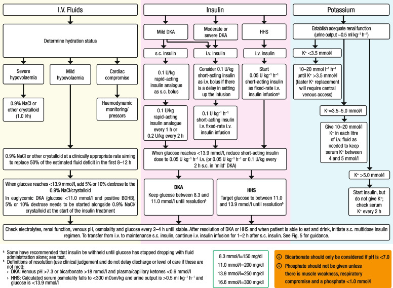
Decision 1: Restore circulation safely
Use an appropriate intravenous crystalloid, such as isotonic saline or a balanced crystalloid where supported by the treatment protocol.
Fluid replacement depends on haemodynamic stability, dehydration, sodium, renal function, cardiac function and response to treatment.
Frailty, kidney impairment and heart failure require particular caution.
Decision 2: Check potassium before insulin
The 2024 international consensus recommends delaying insulin when potassium is below 3.5 mmol/L, while urgently correcting hypokalaemia with monitoring.
Some local protocols use different thresholds or replacement schedules.
OSCE scenario: Potassium is 3.2 mmol/L in a patient with DKA.
The correct response is not to start insulin immediately without addressing potassium. Escalate, commence appropriate monitored potassium replacement and follow the protocol for safely initiating insulin once potassium has been corrected.
This decision can prevent life-threatening arrhythmias.
Decision 3: Understand the difference in insulin strategy
In DKA, insulin suppresses ketogenesis and is essential to resolving ketoacidosis.
Severe DKA generally requires supervised intravenous insulin treatment. Selected uncomplicated mild or moderate cases may be suitable for closely monitored subcutaneous rapid-acting insulin protocols.
In isolated HHS without significant ketoacidosis, fluid replacement takes priority initially. According to the JBDS approach, insulin is generally introduced when glucose and osmolality stop falling appropriately with fluids alone, unless significant ketosis requires earlier insulin.
Where indicated, HHS protocols typically use a lower fixed intravenous insulin rate than DKA protocols.
The precise prescription must follow local adult pathways and clinical supervision.
Decision 4: Avoid rapid osmolality correction in HHS
The recommended target for a gradual fall in osmolality is approximately 3–8 mOsm/kg/hour.
The international consensus also advises avoiding excessively rapid glucose reduction and limiting sodium decline to approximately 10 mmol/L over 24 hours.
These measures help minimise neurological injury and other complications.
Decision 5: Add dextrose when needed
In conventional DKA, glucose may fall before ketones and acidosis resolve.
Dextrose-containing fluids are then introduced according to the protocol so that insulin can continue safely.
Euglycaemic DKA is different: When glucose is already near normal, 5–10% dextrose may be required from the beginning of treatment alongside the appropriate fluids and insulin.
SGLT2 inhibitors should be stopped on admission in patients presenting with SGLT2-associated DKA.
Decision 6: Recognise mixed DKA/HHS
When significant ketoacidosis coexists with hyperosmolality, insulin treatment must address DKA while fluid management accounts for the risk of rapid osmotic changes.
The international consensus recommends DKA-directed intravenous insulin treatment in mixed presentations, with close monitoring and specialist supervision.
Decision 7: Monitor until genuine resolution
Monitoring should include vital signs, mental status, fluid balance, urine output, glucose, ketones, electrolytes and acid–base status at protocol-defined intervals.
According to the international consensus, biochemical DKA resolution requires beta-hydroxybutyrate <0.6 mmol/L together with venous pH ≥7.3 or bicarbonate ≥18 mmol/L.
HHS resolution requires broader assessment of osmolality, corrected hyperglycaemia, restored circulating volume, urine output and neurological recovery.
A normal glucose reading by itself is never sufficient to declare either crisis resolved.
11. Four examiner-style clinical cases
Try answering each case aloud before reading the model response.
Case 1: The patient who missed insulin
A 21-year-old with type 1 diabetes presents with vomiting and deep breathing after missing insulin during an illness.
Glucose 25 mmol/L; beta-hydroxybutyrate 5.6 mmol/L; pH 7.17; bicarbonate 11 mmol/L; potassium 5.1 mmol/L.
Examiner questions
- What is the diagnosis and likely severity?
- Why is the potassium result potentially misleading?
- What are your immediate priorities?
Reveal examiner model answer
The findings indicate moderate DKA. Elevated serum potassium does not exclude total-body potassium depletion. Immediate priorities are ABCDE, escalation, intravenous access, fluid and electrolyte assessment, ECG, supervised DKA treatment, repeated biochemical monitoring and identification of the precipitant.
Case 2: Confusion after several days of illness
A 76-year-old with type 2 diabetes presents with progressive confusion, severe thirst and dehydration.
Glucose 42 mmol/L; sodium 152 mmol/L; urea 16 mmol/L; beta-hydroxybutyrate 0.8 mmol/L; pH 7.37; bicarbonate 23 mmol/L.
Examiner questions
- Calculate effective and total osmolality.
- What is the most likely diagnosis?
- Why might immediate insulin be inappropriate?
Reveal examiner model answer
Effective osmolality is 346 mOsm/kg and total calculated osmolality is 362 mOsm/kg. This supports HHS without significant ketoacidosis. Controlled fluid replacement and monitoring of osmolality, sodium and neurological status are initial priorities. Starting insulin too early may cause unsafe osmotic shifts and circulatory deterioration. Insulin timing follows the local HHS pathway.
Case 3: The deceptively reassuring glucose
A 28-year-old with type 2 diabetes taking empagliflozin develops vomiting, abdominal pain and rapid breathing after poor oral intake.
Glucose 9.8 mmol/L; beta-hydroxybutyrate 4.7 mmol/L; pH 7.22; bicarbonate 13 mmol/L; potassium 4.2 mmol/L.
Examiner questions
- Can this patient have DKA despite glucose below 11.1 mmol/L?
- Why is empagliflozin relevant?
- How does the glucose level affect treatment?
Reveal examiner model answer
This patient has euglycaemic DKA. Established diabetes, significant ketonaemia and metabolic acidosis satisfy the diagnostic criteria. SGLT2 inhibitors can contribute to ketoacidosis without marked hyperglycaemia, especially during reduced intake or illness. Stop the SGLT2 inhibitor, escalate, initiate the supervised DKA pathway, monitor potassium and ensure dextrose is provided appropriately from the outset so that necessary insulin treatment can continue safely.
Case 4: The mixed emergency
A 67-year-old with type 2 diabetes presents with profound dehydration, drowsiness and vomiting following an infection.
Glucose 39 mmol/L; sodium 151 mmol/L; urea 16 mmol/L; beta-hydroxybutyrate 5.2 mmol/L; pH 7.16; bicarbonate 12 mmol/L.
Examiner questions
- Calculate effective osmolality.
- Is this uncomplicated HHS?
- Which management principle must not be missed?
Reveal examiner model answer
Effective osmolality is (2 × 151) + 39 = 341 mOsm/kg. Total calculated osmolality is 357 mOsm/kg.
The significant ketonaemia and acidosis indicate DKA, while the substantial hyperosmolality indicates overlapping HHS features.
This is a mixed DKA/HHS presentation.
The patient requires urgent senior-led management that treats ketoacidosis with appropriate insulin while avoiding excessive osmotic changes during fluid replacement. Potassium, clinical status and biochemical trends require close monitoring.
12. How to present your findings to an OSCE examiner
A structured clinical presentation should communicate four things:
Diagnosis → Supporting evidence → Immediate danger → Management plan
Example: DKA
“This patient has diabetic ketoacidosis, supported by significant ketonaemia and metabolic acidosis in the context of diabetes. I am particularly concerned about dehydration and potassium disturbances. I would urgently escalate, perform an ABCDE assessment, establish intravenous access, check electrolytes and ECG, and initiate the appropriate DKA pathway under senior supervision. I would monitor ketone clearance, acid–base status and the clinical response while investigating the precipitating cause.”
Example: HHS
“This patient has likely hyperosmolar hyperglycaemic state, demonstrated by severe hyperglycaemia, marked hyperosmolality and dehydration without significant ketoacidosis. I would urgently request senior review, assess ABCDE, establish intravenous access and begin carefully monitored fluid replacement. I would follow the HHS pathway for insulin timing, monitor osmolality and electrolytes closely, and investigate infection or other precipitating illness.”
For additional examiner-focused preparation, see OSCE.ai's guide to OSCE mark schemes.
13. Patient counselling and prevention of recurrence
Preventing another diabetic emergency is an important communication task.
Once the patient is medically stable, explore the cause of the episode without blame.
Review insulin access, missed doses, injection technique, pump function, medications, glucose and ketone monitoring, and the patient's understanding of illness management.
Patients using insulin should receive an individualised sick-day plan.
Explain that basal insulin must not simply be stopped during illness, even when food intake is reduced. Dose adjustments and additional monitoring should follow their established diabetes-team instructions.
Patients should know when to test ketones, when to contact their diabetes team and when persistent vomiting, rising ketones, breathlessness, confusion or inability to maintain fluids requires urgent medical assessment.
Discuss temporary interruption of medicines such as SGLT2 inhibitors during relevant acute illnesses according to individualised guidance. Following SGLT2-associated DKA, any consideration of restarting treatment requires specialist risk–benefit assessment.
Use plain language, confirm understanding and arrange appropriate follow-up.
14. OSCE self-assessment: A 20-point practice checklist
Use this educational rubric after a timed practice station. It is a proposed self-assessment tool, not an official university marking scheme.
| Performance domain | Marks |
|---|---|
| Recognises DKA, HHS or mixed crisis and states urgency | 2 |
| Calls for help and describes a safe ABCDE assessment | 3 |
| Requests and interprets appropriate investigations | 4 |
| Explains fluid, potassium and insulin safety priorities | 4 |
| Includes monitoring, reassessment and precipitant management | 3 |
| Communicates reasoning clearly and provides a safe handover | 2 |
| Addresses resolution, prevention and follow-up | 2 |
| Total | 20 |
Safety-critical omissions include failing to recognise deterioration, neglecting potassium before insulin, treating euglycaemic DKA as reassuring, or applying an inappropriate rapid-correction strategy to HHS.
For a structured method of practising clinical stations, visit How to Practice OSCE Stations.
15. Frequently asked OSCE questions
Can DKA occur with normal or mildly elevated glucose?
Yes. Euglycaemic DKA occurs when DKA-associated ketosis and acidosis are present with glucose <11.1 mmol/L. SGLT2 inhibitor treatment, reduced food intake and other clinical circumstances are important clues.
Why is blood beta-hydroxybutyrate preferred?
It directly measures the predominant ketone body in DKA, improving diagnosis and monitoring compared with conventional urine ketone tests.
Is potassium always low in DKA?
No. Serum potassium may initially be normal or elevated despite total-body depletion. It can fall dangerously after insulin treatment begins.
Why shouldn't HHS be treated exactly like DKA?
Because severe hyperosmolality and volume depletion require careful correction. In isolated HHS, initial fluids and controlled osmolality reduction are particularly important, and insulin timing differs.
What indicates DKA has resolved?
Under the international consensus, beta-hydroxybutyrate <0.6 mmol/L together with venous pH ≥7.3 or bicarbonate ≥18 mmol/L indicates biochemical resolution. Clinical stability and an appropriate insulin transition remain essential.
What is the most important OSCE lesson?
Interpret the biochemical pattern, recognise the dangerous physiology, and explain the next safe clinical action.
Final OSCE takeaway
Before finishing a DKA or HHS station, ask yourself:
Have I recognised the emergency? Have I assessed ABCDE and requested help? Have I interpreted glucose, ketones, pH, bicarbonate, osmolality and potassium correctly? Have I explained the safest treatment priorities and how I will monitor the response?
The highest-value clinical skill is not merely remembering a numerical threshold. It is understanding how each result changes what you should do next.
Strengthen that skill by practising unfamiliar presentations, answering examiner questions aloud and reviewing mistakes after every station.
Explore more learning resources in the OSCE.ai Resources Library and practice with 200+ OSCE stations.
References and further reading
- American Diabetes Association Professional Practice Committee for Diabetes. Diabetes Care in the Hospital: Standards of Care in Diabetes—2026. Diabetes Care. 2026;49(Suppl 1).
- Umpierrez GE, Davis GM, ElSayed NA, et al. Hyperglycemic Crises in Adults With Diabetes: A Consensus Report. Diabetes Care. 2024;47(8):1257–1275.
- Joint British Diabetes Societies for Inpatient Care. The Management of Diabetic Ketoacidosis in Adults. Revised March 2023.
- Mustafa OG, Haq M, Dashora U, Castro E, Dhatariya KK. Management of Hyperosmolar Hyperglycaemic State in Adults: An Updated JBDS Guideline. Diabetic Medicine. 2023;40(3):e15005.
- Feingold KR, Dhatariya K. Diabetic Ketoacidosis. Endotext. Updated January 2026.
- Dhatariya K, Mustafa O, Stathi D. Hyperglycemic Crises. Endotext. Updated June 2025.
- Resuscitation Council UK. The ABCDE Approach. Updated July 2024.
- Chiasson JL, Aris-Jilwan N, Bélanger R, et al. Diagnosis and treatment of diabetic ketoacidosis and the hyperglycemic hyperosmolar state. CMAJ. 2003;168(7):859–866. Original pathophysiology diagram, Fig. 1.
- Rogers J, Cadogan M. Blood Gas Interpretation. Life in the Fast Lane. Reference acid–base flowchart.
Educational scope: Adult OSCE preparation. This article is not a bedside prescribing protocol. Follow locally approved emergency pathways and specialist supervision. Independent sign-off by an appropriately qualified clinical reviewer is recommended before publication.